Table of contents
WHAT IS THE MACULA?
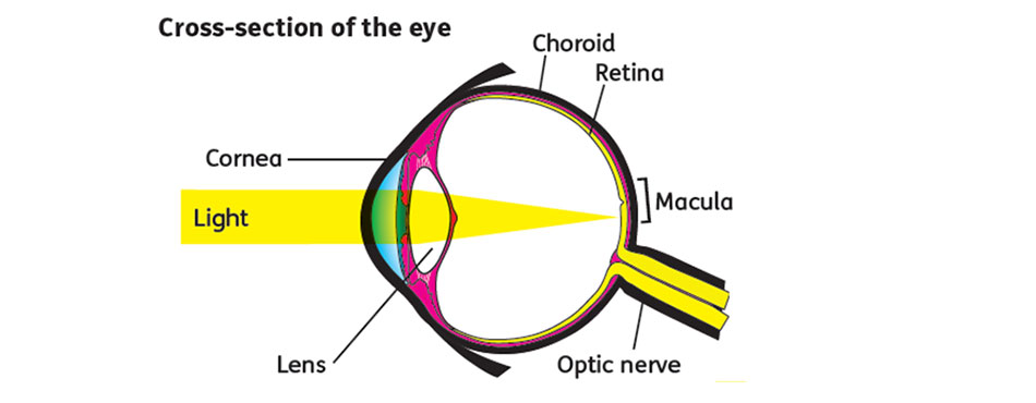
The macula is part of the retina at the back of the eye. It is only about 5mm across but is responsible for our central vision, most of our colour vision and the fine detail of what we see.
The macula has a very high concentration of photoreceptor cells – the cells that detect light. They send signals to the brain, which interprets them as images. The rest of the retina processes our peripheral, or side vision.

WHAT IS AGE-RELATED MACULAR DEGENERATION?
Age-related Macular Degeneration (AMD) usually affects people over 60 but can happen earlier. The older we are the greater our risk of developing the condition. Around one in every 200 people has AMD at 60. However by the age of 90 it affects one person in five. We are all living longer so the number of people affected is increasing. AMD is the most common cause of sight loss in the developed world.
There are two forms of AMD:
- Dry AMD is a gradual deterioration of the macula as the retinal cells die off and are not renewed. The progression of dry AMD varies but in most people it develops over many months or years. Often people carry on as normal for some time. There is currently no treatment for dry AMD.
- Wet AMD develops when abnormal blood vessels grow into the macula. These leak blood or fluid which leads to scarring of the macula and rapid loss of central vision. Wet AMD can develop very suddenly. It can now be treated if caught quickly.
Around 10 to 15 per cent of people with dry AMD develop wet AMD so if you have been diagnosed with the dry form of the disease and notice a sudden change in your vision, contact your optometrist or hospital eye specialist urgently.
If you have AMD in one eye, the other eye may be affected within a few years.
SYMPTOMS
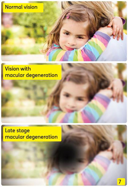
Macular degeneration affects people in different ways. Symptoms may develop slowly if you have dry AMD, especially if it’s only in one eye. However, as the condition progresses, your ability to see clearly will change.
- Gaps or dark spots (like a smudge on glasses) may appear in your vision, especially first thing in the morning.
- Objects in front of you might change shape, size or colour or seem to move or disappear.
- Colours can fade.
- You may find bright light glaring and uncomfortable or find it difficult to adapt when moving from dark to light environments.
- Words might disappear when you are reading.
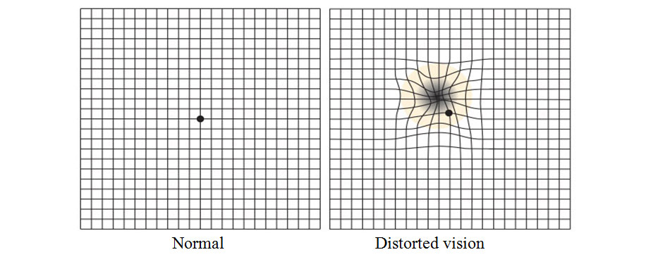
- Straight lines such as door frames and lampposts may appear distorted or bent.

DIAGNOSIS
Your ophthalmologist will ask questions regarding recent changes to your central vision and will perform a complete eye exam.
- Amsler grid, which is a patterned image that looks like a checkerboard. Early changes in your central vision will cause the grid to appear distorted

- Eye drops will be placed in your eyes to enlarge (dilate) your pupils. Dilating the pupils allows your ophthalmologist to view the back of the eye clearly.
- Fluorescein dye angiography: A dye injected into a vein in the arm travels to the eye, highlighting the blood vessels in the retina so they can be photographed. The dye will temporarily change the colour of your urine, so be prepared.
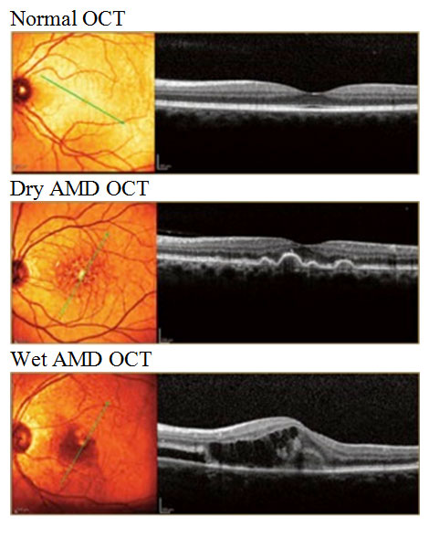
- Ocular Coherence Tomography (OCT): non-invasive test, similar to an ultrasound, which creates cross-sectional images of the retina and can detect fluid within the macula.

RISK FACTORS FOR AMD
The cause of AMD is not known but there are a number of factors associated with the development of the condition.
- Age is the main risk factor. As we age, cell regeneration reduces. This increases the risk of developing the condition.
- A family history of macular degeneration will increase your chances of developing AMD.
- Smoking damages blood vessels and the structure of the eye. Smokers are up to four times more likely to develop macular degeneration than non-smokers. If you also have the particular gene for AMD you are twenty times more likely to develop the condition.
- A poor diet low in fruit and vegetables may increase the risk of AMD. Antioxidants and other substances in fruit and vegetables protect the body against the effects of free radicals. These are unstable molecules that damage cells or prevent cell repair.
- Alcohol destroys antioxidants and is therefore a risk factor.
- Obesity and a diet with lots of sugars and hydrogenated or saturated fats also increase the risk of developing AMD.
- People with high blood pressure are one and a half times more likely to have AMD than those with normal blood pressure.
- Sunlight: macular cells are sensitive to the Ultra Violet (UV) radiation and visible blue light, which occurs naturally in sunlight. Cell damage from blue light may cause deterioration of the macula.
TREATMENT
Treatment of dry AMD
There is currently no treatment to cure dry AMD. The treatment for early dry AMD is generally nutritional therapy, with a healthy diet high in antioxidants to support the cells of the macula. If AMD is further advanced but still dry, supplements are prescribed, to add higher quantities of certain vitamins and minerals which may increase healthy pigments and support cell structure (Vitamins C and E, beta carotene, zinc, and antioxidants).
Lifestyle changes may help protect your eyes:
- Do not smoke. This is the most important self-help measure you can take. If you would like help to stop smoking Take an appointment at the Quit Smoking Clinic of FV Hospital;
- Maintain a healthy weight and blood pressure;
- Eat a diet low in saturated fats and rich in omega 3 fatty acids (e.g. oily fish, walnuts) and fresh fruit and vegetables;
- Don’t drink alcohol to excess;
- Take regular exercise;
- Wear lenses that block UV and blue light, and reduce glare;
- Wear a hat with a brim or visor to shade eyes from direct sunlight;
- Have regular eye tests to spot problems;
- Monitor your vision to check for changes.
Treatment of wet AMD
- Anti-VEGF therapy
- VEGF is an acronym for Vascular Endothelial Growth Factor. Currently, the most common and effective clinical treatment for wet AMD is anti-VEGF therapy – which is periodic intra-vitreal (into the eye) injection of a chemical called an “anti-VEGF”
- This treatment helps reduce the number of abnormal blood vessels in your retina. It also slows any leaking from blood vessels.
- An injection in the eye can be a disconcerting experience, and it may take several treatments to become accustomed to the procedure. However, the shot is usually not painful because the eye has been anaesthetised. The procedure takes about fifteen minutes. Usually the appointment requires an hour. The effect lasts for a month or maybe more. You usually need a course of monthly injections over a year or longer.
- Laser surgery may also be used to treat some types of wet AMD. Your ophthalmologist shines a laser light beam on the abnormal blood vessels. This reduces the number of vessels and slows their leaking.


