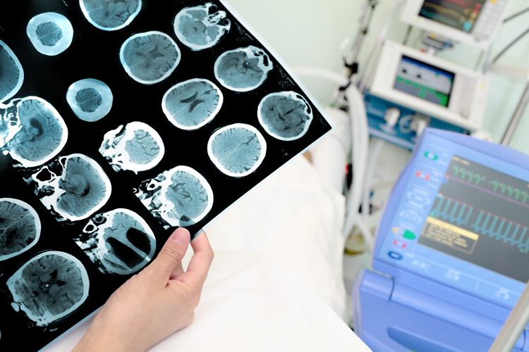
FVH’s Imaging Department plays an important role in providing diagnostic imaging services to meet the demands of patients examined and treated at all departments of the hospital. X-ray, ultrasound, computerised tomography (CT) scans and Magnetic Resonance Imaging (MRI) provide detailed images of all parts of the body, such as the brain, cardiovascular system, internal organs, bones, joints, blood vessels, spinal marrow and breasts.
FVH’s Imaging Department applies state-of-the-art technologies to provide precise diagnostic results quickly and is key in helping to diagnose complicated diseases which cannot be diagnosed by other disciplines. The department’s strengths include:
1. A team of highly skilled and experienced radiologists and imaging technicians
Each member of the Imaging Department radiologist team has completed advanced training courses in imaging in France, and FV regularly sends radiologists abroad to pursue fellowships and participate in intensive training courses in order to continuously upgrade their skills and ensure that the latest imaging technologies are offered at FVH. Additionally, we also collaborate with imaging specialists from France and the team of Magnetic Resonance Imaging (MRI) specialists at the Medical University Hospital of Ho Chi Minh City. Our imaging technicians are also trained intensively by GE specialists from Singapore and Indonesia so that they can operate the Imaging Department’s sophisticated equipment in accordance with international protocols.
2. New and advanced technologies and state-of-the-art equipment.
FVH has an MRI scan which can support the detection and diagnosis of the smallest injuries in our body, such as damage to the blood vessels, brain, spine and joints. The CT scanner can image the organs of the body through arthroscans, angioscans, dentascan, CT colonography (also known as virtual colonoscopy, using for the screening of colon cancer) and other specialised scans; mammography for the early detection of breast cancer; colour Doppler; X-ray scans, and so on. All of these scans provide accurate results.
At present, FVH’s Imaging Department is equipped with an integrated machinery system manufactured by GE (USA), including:
- Five X-ray scanners, including two X-ray scanners, one digital X-ray scanner, one maxillofacial X-ray scanner and one mobile X-ray scanner for bed-ridden patients in emergency and surgery cases.
- Mammography scanner
- Colour ultrasound scanner
- A new GE BrightSpeed Elite CT Scanner for fast image acquisition, wide coverage, high definition and clear images.
- MRI Optima MR360 1.5T scanner, which uses a magnetic field and radio waves to produce very detailed, high-quality images of parts of the body in very little time. This procedure is extremely safe for patients to undergo as no X-rays are used.
3. Multidisciplinary consultations
FVH is a general hospital and its Imaging Department plays an extremely important role in patient care. The Imaging Department performs imaging investigations for all of FV Hospital’s departments, including Otolaryngology, Gastroenterology and Hepatology, Urology and Andrology, Paediatrics, Orthopaedics and Neurosurgery. Once imaging tests have detected any health conditions that require the attention of specialties in the hospital, doctors from each will participate in a multi-disciplinary consultation to develop the safest and most effective treatment plan. This ensures that treatment costs and impositions on the patient’s time are minimised.
4. Impeccable hygiene standards and full patient protection during every investigation and examination
The FVH Imaging Department offers a wide variety of services and treatments, at all times maintaining high standards of hygiene and protection for patients during every investigation and invasive examination. For examples, when an X-ray scan is performed on one area of the body, the rest of the patient’s body will be covered by a lead glass shield to protect it from radiation.
5. Services for referred patients
In addition to providing services for patients at FVH, the Imaging Department also provides services for patients referred by other international clinics in HCMC. Patients who are referred for special medical tests can book an appointment with a physician via the telephone number (028) 5411 3400.
X-ray Scanner
AnX-ray scan is a commonly used medical imaging procedure which plays an important role in the diagnosis and treatment of diseases.
X-ray scans are performed when requested by doctors to screen for diseases and injuries, for periodic health examinations, recruitment health examinations, and so on. An X-ray scan takes around 15 minute to complete and in most cases no preparation is required.

However, when the stomach, oesophagus, duodenum and colon are scanned, prior dietary and bowel preparations may be required.
During the X-ray scan, you should follow the instructions of doctors or physicians to ensure the best results are obtained. You should notify your doctor or physician if you are pregnant or think there is a possibility that you may be pregnant.
FVH’s Imaging Department provides the following X-ray scans:
- Conventional radiography for lungs, sinuses, skull, spine and bones.
- Digital fluoroscopy for the examination of the gastrointestinal tract, uterus and oviducts with contrast, studies of the urinary system, arthroscopy, percutaneous biopsy and drainage.
Mammography (Breast Imaging)
Mammography, often called breast imaging, is used for the early detection and diagnosis of breast abnormalities. At FVH, we usually offer breast cancer screening programmes to patients that require this procedure. Women aged from 35 to 40 years should periodically undergo mammography, ideally once or twice a year.
Our state-of-the-art mammography equipment allows us to perform full field mammography and stereotaxic core biopsies by applying a three-dimensional location technique. This technique gives accurate results and minimises omissions.
During breast imaging, the patient’s breast will be placed on a plate and tightly compressed with a plastic paddle. This breast compression facilitates the detection of abnormalities, including small lesions, expands the field of observation and increases image quality.
The breast cannot be positioned so that a complete single scan can be taken. Breast imaging is performed in several positions to obtain all the necessary images of the breast:
- Top-to-bottom view: the X-rays will pass from the top to the bottom through the breast.
- Side-to-side view: the X-rays will pass from one side to the other though the breast, from left to right or vice versa depending on whether the left breast or right breast is being scanned.
- Angle-side view: the X-rays will pass through the breast at an angle
Ultrasound Scanning
Ultrasound scanning is a safe imaging method which poses no threat to the patient’s health. FVH prints ultrasound results on film rather than paper to increase the longevity and integrity of the printed image.
Patients undergo ultrasound scans for routine disease screening or when one is requested by a clinical doctor. Ultrasound is also used to continually follow the development of a disease. During an ultrasound scan, a layer of harmless gel is applied to the skin to help the ultrasound waves to penetrate the skin and tissues underneath. At FVH, we offer the following ultrasound scans:
- Brain
- Parotid glands
- Breast
- Thyroid, including neck lymph nodes
- Neck and under-chin area
- Cheek
- Armpit
- Groin area
- Abdominal area
- Kidney and bladder
- Pelvic area
- Testicular
- Prostate
- Spinal
- Musculoskeletal
- Soft parts of the body
In addition to the above services, a colour Doppler ultrasound permits the study of blood flows inside arteries and veins. We also apply ultrasound in biopsy positioning.
CT Scans (Computerised Tomography)
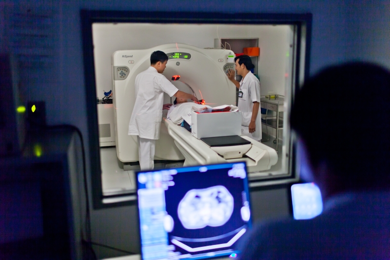
This is an imaging technique using X-rays in combination with a multi-row detector in order to create detailed images inside the body in a very short time.
Generally, you do not need to prepare for a CT scan, although some specialised CT screens may require the injection of contrast dye through the vein.
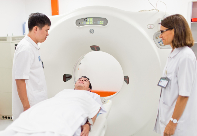
During the scan, you should follow the instructions given by the technician and remain completely still. You should notify the doctor and technician if you are pregnant or if there is a chance you may be pregnant, or have diabetes or a kidney-related disease. FVH’s Imaging Department offers the following CT scans:
- Parts of the body such as the skull, facial bones, ear, neck, molars, abdominal area, pelvic area, genitourinary apparatus, spine, hip, chest, sinus and orbit; arthroscan with contrast.
- Dentascans and other specialist scans.
- Vascular imaging, mainly of arteries such as the coronary artery, peripheral artery, aorta, pulmonary artery and renal artery.
- CT colonography for intestinal analysis by inflating the colon with carbonic gas through an automatic pump. FV’s Imaging Department is the only place to offer this procedure in Vietnam.
FV also offers interventional procedures such a CT scanner-guided biopsies and drainage.
Magnetic Resonance Imaging (MRI)
This is the latest technology to create images of the inside of the body. MRI scans use strong magnetic fields and radio waves to produce a detailed image of the organs, tendons and bone, tissue, and brain. Images can be transferred to film (hard copy) or recorded on CD or DVD (soft copy).
FVH’s Imaging Department utilises the Optima™ MR360 1.5T, which combines the latest technology with dedicated software to produce high-quality images in a short period of time. Doctors will use these images to evaluate parts of the body and reach an accurate diagnosis.
One of the main advantages of MRI is that it uses no X-rays and therefore does not expose the body to radiation. The Optima™ MR360 1.5T is also one of the most patient-friendly MRI scanners, designed to ensure that patients feel comfortable and completely at ease. In almost all MRI cases, there is no special preparation required and patients simply have to dress comfortably when visiting the hospital for an MRI procedure.
When an MRI scan is performed, the patient lies in the bore of the magnet within a very strong magnetic field, which means any implanted metal devices in your body may be affected. This is why MRI scans require a prescription from a doctor. You must remain relaxed and completely still during the scan so that the highest quality images can be obtained. Typically, an MRI exam will take approximately 30 – 45 minutes per area scanned, depending on the information that the doctor requires for diagnosis.
You should inform the radiologists or radiographers if you have any of the following:
- A pacemaker
- Aneurysm clips
- Metal fragments in your eye
- Any other implants under the skin (for patients with diabetics)
- Any other metal implants (surgical staples, artificial teeth, artificial joints, etc)
- If you think you are pregnant.
You must remove items like your jewellery, wallet and cards with a magnetic strip (such as ATM and credit cards), watch, keys, hair pins and phone before stepping into the MRI room. Bring along your medical records and prior screening results (X-ray films, ultrasound results, CT scans and MRI scan results, etc.).
MRI services at FVH’s Imaging Department:
- Musculoskeletal imaging, i.e. imaging of all the joints, tendons, bones and tissues of superior and inferior extremities such as the wrist, arm, elbow, hand, hip, thigh, knee, shin, ankle, and foot.
- Neurological imaging, i.e. imaging of the skull, brain, cerebral vessels and spinal cord.
- Head and neck imaging, i.e. imaging of the neck spine, ear cavity, orbit, nasal cavity, larynx, salivary glands, lymph nodes, pituitary gland.
- Imaging of the chest wall, thoracic spine, thoracic aorta.
- Lumbar spine imaging.
- Abdomen and pelvis imaging, i.e. imaging of the abdomen and abdominal wall, bile duct, pancreas, urinary system, uterus, cervix and ovaries (female), prostate and testicles (male), anal canal, pelvis floor, etc.
- Biliary and pancreatic duct imaging.
- Vascular imaging (imaging carotid artery, cerebral blood vessels, aorta, internal arteries, extremity arteries).
- Breast imaging.
In addition to providing imaging services within FVH’s Imaging Department, we also offer services at two other locations for our patients’ convenience:
Mobile imaging services, such as on-bed X-ray scans for patients in FV Hospital’s Emergency Department, Intensive Care Unit (ICU) and Operation Theatre (OT).
X-ray scans and ultrasound scans at FV Saigon Clinic, Third floor, Bitexco Financial Tower, 2 Hai Trieu Street, District 1, HCMC.

FVH is equipped with the latest radiology technology and is the General Electric Medical Reference Centre for Vietnam. All our equipment is Dicom compatible and all films are digitalised using the Agfa Centricity System.
Our inventory of state-of-the-art equipment includes:
Proteus X-ray units for conventional radiography
A Prestige II remote-controlled table, installed in the scan room along with fluorescent lights and a TV system. This device enables the technician to target the patient’s body parts that need screening via remote control. The digital X-ray system allows computer processing of images and results in high quality images which can be zoomed in or zoomed out on the areas under observation. Contrast can also be adjusted.
An Orthoralix 9200 X-ray panoramic radiography system, used to screen one-dimensional perspectives of teeth and bone structure. This is a safe and convenient procedure that takes a very short time and provides high quality images.
A Mobile X-ray system, used to perform X-ray scans on bedridden patients. A Senographe DMR+ mammograph system, equipped with three-dimensional location technology and a special light to read mammographic film.
A Siemens Sonaline G60S Logic 5 digital calibrated Doppler ultrasound system, which provides detailed, high quality images which support accurate diagnosis and minimise the possibility of needing to do the procedure again.
A BrightSpeed Elite CT scanner, which provides improved images and faster image acquisition when compared to other machines, as well as virtual image dissection. The new CT-scanner produces clearer CT images of the vascular system including the main arteries in the neck, the peripheral artery, aorta, pulmonary artery and renal artery and effectively assists the detection of arteriostenosis. Additionally, this machine can be used to analyse the large intestine via CT colonography to identify protuberance and polyps located within the organ. The high image quality produced by this machine is vital to doctors when seeking a more accurate diagnosis.
A new GE OptimaTM MR360 1.5T MRI scanner, which combines the latest technology with dedicated software to produce high-quality images in a short period of time. It is also one of the most patient-friendly MRI scanners, designed to ensure that patients feel comfortable and completely at ease. The versatile Optima™ MR360 is an extremely useful tool in nearly any clinical situation, ranging from routine orthopaedic exams to complex breast, body, or neurological cases.
No results found...


